MIM Symphony Bx


| MR Guided Biopsy |
| without an MR Suite |
The new standard in prostate imaging for cancer detection is multi-parametric MR (T2W, DCE, DWI). Until now, urologists have not been able to effectively use this targeted information to guide their biopsy sampling. MIM Symphony Bx is changing this with powerful yet easy-to-use tools for everyone on the MR guided biopsy team.

View the following 13 minute video to see how MIM Software and Nuada Medical are working to improve MR-US fusion for prostate diagnostics and biopsy.
"Symphony Bx has outstanding capabilities in integrating multiple imaging tools in the diagnosis and treatment of prostate cancer. We use it to easily plan MR-targeted biopsies, design transperineal mapping, plan brachytherapy seed implants, and aid in cryotherapy salvage. It is an amazing advance in prostate cancer management."
Peter Grimm
DO
Prostate Cancer Center of Seattle
Seattle, Washington
Prostate Cancer Center of Seattle
Seattle, Washington
MR

The new standard in prostate imaging for cancer detection is multi-parametric MR (T2W, DCE, DWI). Until now, urologists have not been able to effectively use this targeted information to guide their biopsy sampling. MIM Symphony Bx is changing this with powerful but easy-to-use tools for everyone on the MR guided biopsy team.
Radiology

MIM provides advanced diagnostic tools for MR, including the ability to display and fuse multi-parametric MR images. Suspicious lesions can be contoured on any plane or sequence and are dynamically displayed on each MR image. Time activity curves are automatically generated to highlight wash-in and wash-out of contrast. With Reslicer™, the radiologist or urologist can prepare the contours for use with TRUS-guided biopsy by reorienting the MR to match the TRUS images – all before the patient arrives.
Urology

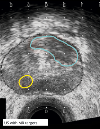
At the start of the procedure, the urologist is only a few clicks away from displaying the radiologist-defined MR targets on the live TRUS image. Using a point-and-shoot approach, the targets can be sampled and the precise locations of the samples recorded. With little additional time and effort in the room, biopsies can now be focused on areas of high suspicion as identified by MR.
Pathology

MR guided biopsies provide the potential for higher yield and more accurate staging. Nobody benefits more from correlating the final pathology results with imaging findings than the patient. However, the urologist can be more confident in patient management and treatment decisions and the radiologist more accurate with follow-up and future imaging.
MIMcloud

MIMcloud facilitates this groundbreaking collaboration between radiologist and urologist. This Internet-based service ensures on-demand access to MR images, outlined contours, and reports – every step of the way. If a repeat MR is ordered, the previous images, contours, and biopsy records are retained for comparison.


 京公网安备11010802039423号